There has never been a better time to be a refractive surgery patient than right now. The advancements in both cornea-based and lens-based refractive surgery make it possible to provide a solution for nearly every patient seeking vision correction.
There has also never been a better time to be a refractive surgery practice in terms of the diagnostic and surgical tools available to us.
I have had the opportunity to serve several roles in my refractive surgery career that spans more than two decades. I currently serve as director of refractive surgery for Rush Eye Associates in Amarillo, TX, while also serving as a consultant to other practices and to the ophthalmic industry. In these roles, I’m always looking for a way to improve efficiencies, enhance our decision-making process, and improve our patient outcomes.
Below, I will share with you how I use diagnostic technology, particularly wavefront technology, during my preoperative patient education along with how the devices help me with decision-making to determine which refractive surgery may suit them the best.
Wavefront devices
Wavefront diagnostic devices have evolved over the last two decades. Not only can we use these devices to measure the entire refractive status of the eye, but we can also identify where the optical imperfections are originating from. There are different types of wavefront devices, such as ray tracing, Hartmann-Shack, and dynamic sciascopy, among others.
My practice uses three different wavefront diagnostic devices in our refractive surgery work-ups: the Marco OPD-III, the Visionix VX-120, and the Tracey Technologies iTrace. I use each of these devices several times per day, and many patients have all three diagnostic devices performed on them during the visit. Each of these devices measures the optical system in a slightly different way.
The OPD-III is a wavefront aberrometer that provides refraction and topography results. It also provides pupil size in various lighting conditions and gives the provider a retroillumination image.
The VX-120 features the combined functions of an autorefractor, a keratometer, a corneal topographer, a wavefront aberrometer, a pachymeter, and a non-contact tonometer along with anterior chamber analysis.
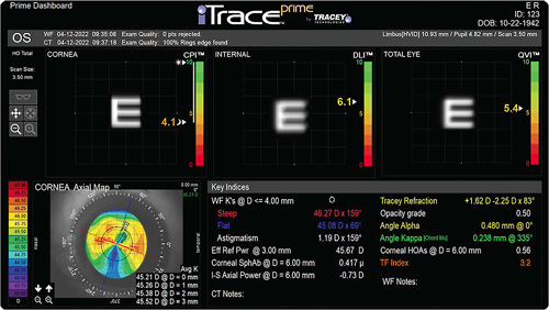
The iTrace provides a visual representation of the quality of vision associated with the cornea, the internal workings of the eye, and the sum of both. There is also a numerical analog score on a scale of 1-10, with 10/10 representing pristine optics and 1/10 representing optics that have some type of degradation in them.
Case study
Here is an example of a patient that I recently worked with in our clinic. The female patient was a 45-year-old registered nurse who was interested primarily in LASIK when she presented.
Some practices will immediately lean towards performing lens-based refractive surgery, such as refractive lens exchange, dysfunctional lens replacement, or cataract surgery, on any patient that is in the presbyopic age range. In our practice, we take the time to discuss cornea- and lens-based options with the patient while using our diagnostics to explain our reasoning.
At first glance, our patient was a pretty good LASIK candidate. She had excellent corneal topography, pachymetry, and her BCVA was 20/20 in each eye. She didn’t mind wearing reading glasses. However, I happen to notice that my manifest refraction showed a slight increase in her myopic refractive error. Because of this, I ordered an iTrace on the patient, which we use to help determine which refractive surgery procedure to recommend, while also combining its data and that of other diagnostic devices (including the IOLMaster [Zeiss] and Lenstar [Haag-Streit] or Argos [Alcon] biometers) in the surgical planning of the recommended procedure.
Even though the patient’s slit lamp examination of the lens was unremarkable, the iTrace picked up a deterioration of the internal optics. I further quizzed the patient about her quality of vision, and she stated she had difficulty driving at night. This demonstrates the importance of asking questions as an adjunct to the slit lamp exam, because this information would not have been revealed at the slit lamp. We now know that there are physiological changes that happen in the crystalline lens that manifest even before they are necessarily visible at the slit lamp.
With the iTrace, I was able to show the patient where the deterioration in her vision was coming from using iTrace’s new display, iTrace Prime Dashboard, which allows me to quickly obtain an understanding of the patient’s optical system in a single display (see Figure). This gave her even more confidence in our practice and in my recommendation of lens-based surgery.

Ultimately, we decided on a lens-based refractive surgery. This patient now has an internal optics of 10/10 and is very happy with her vision.
Summary
When deciding on a diagnostic investment for your practice, two things must be true: the investment must be good for your patients, and it must be good for your practice. If the investment assists in enhancing the outcomes of your patients, then, by default, it will absolutely be good for your practice. In addition, no matter which technology you choose to implement into your refractive surgery work-up, it is important to understand the strengths, weaknesses, and tendencies of the technology OP


