Practice
Everyday Tips for Technicians
Simple “light” techniques can improve patient care.
By Josef Tamory, Herndon, Va
Penlight Thumb Technique
Have you ever had difficulty when checking light-colored pupil size in bright light situations or dark pupils in dim light situations?
Dim disposable penlights designed to manage this situation seem to get dimmer by the day. A slightly better solution is to adjust the muscle light to a low level to get the least output. That way, a patient’s pupil does not constrict as much. Or you can try a method that I call the “Tamory Thumb Technique.”
First, dim the exam room light so that the pupils in both light and dark eyes can dilate naturally to their nighttime extent, or indoors (this can also be helpful in LASIK candidates who need to be properly evaluated for natural pupil size during dim or ambient lighting situations). Then, place your thumb right in front of the penlight or muscle light to totally cover the light source. This may seem odd, but in my experience, it works wonders.
Turn on your light source with your thumb covering up the bulb. You now have a nice, soft, red glow that can be placed near the pupil, which will illuminate enough for the pupil size to be evaluated without making the pupil constrict or react too much. When you see things under a red light, it doesn’t degrade the eyes night-vision as much as seeing things under multi-spectrum light. This way, the pupil does not constrict as much as under a “white” light and you also don’t “blind” the patient as much.
The reason this works is due to the different structures within the retina and their designated functions. The retina is made of two types of receptor cells: cones and rods. The cones are responsible for normal daytime vision and detect both the wavelength (color) and the intensity (brightness) of light. Rods are responsible for our “night adapted vision.” Rods do not detect wavelength, but are very sensitive to intensity of light. They only work at very low light intensities (dim light), are most sensitive to light at about 500nm (turquoise/cyan), and are blind to red light (around 620nm).
Fixation Lights
Have you ever performed biometry or keratometry on a patient who was unable to focus on the fixation target or been asked to help a coworker who was experiencing that difficulty? The small red light on the biometry probe appears larger to the patient; the white light in the keratometer often goes undetected.
If you haven’t already noticed how some patients still look around even after you give specific instructions to look only at the fixation target, you have not done enough A-scans. Patients undergoing an A-scan biometry usually have some kind of cataract and their view of the world can be hazy, blurry, foggy, smeared, or bothered by the glare of light. This translates to the little red dot (or whatever the fixation light is) being a big blur. The patient may not be able to correctly identify the direction from which it is coming. This can be confusing to the technician observing the patient’s eyes moving around.
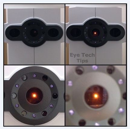
What works for me when performing contact biometry is to simply tell the patient to look at the center of the light. The fixation may seem like a big blur, but ask the patient to picture where the center of that light would be and to keep looking right at it. As the fixation comes closer to the eye, it appears larger (Figure 1). The fixation can also be enlarged or distorted due any opacity the patient may have (for instance, a corneal scar, or cataract). The same goes for the immersion technique. That barrier of fluid (BBS) is just like opening your eyes underwater, which can distort the fixation even with mild cataracts. With the patient prone for an immersion scan, you may want the patient to fixate on a large target affixed to the ceiling to prevent convergence. The same effect can also be seen with the IOL Master’s built in fixation light (Figure 2).

Figure 1: The fixation light on this biometery probe may appear small to the technician, but will appear quite large to the patient. Notice how it becomes larger and larger as it gets closer to the eye. Now picture what it may look like if the patient had a dense cataract.

Figure 2: Here is the fixation light of an IOL Master showing the same effect of how a small fixation light can become very large and distorted to a patient.
These measurements are very important since the slightest movement on the part of the patient looking away from the fixation can result in an incorrect axial length measurement. The slightest movement on the part of the patient looking away from the fixation can result in an incorrect axial length measurement. A measurement that is either too long or shorter than the actual axial length will result in a considerable difference in the post-op refraction. Providing the patient with clear instructions can help the patient and the technician achieve an accurate biometry measurement.
When the patient is unable to fixate in the manual keratometer, use the transilluminator or penlight to help the patient locate the area of fixation. Shine the lit stimulus in the eye piece to catch the patient’s eye. Once central, remove the light, instructing the patient to maintain the fixation.
Because these two measurements are used to determine the power of the intraocular lens, the surgical outcome will be affected if either the corneal curvature or axial length is incorrect. Patient fixation is key when performing various diagnostic tests. Unlike many others, however, these cannot be repeated once the surgery has been performed. OP

|
Josef Tamory is JCAHPO-certified and an ATPO member, also member of the CLAO. He is the founder and creator of Eye Tech Tips on social media sites, and Co-Chair at the ATPO Marketing and Membership Committee. Contact Mr. Tamory by email at eyetechtips@gmail.com or by visiting the website www.facebook.com/eyetechtips. |


